
Local researchers have created an innovative imaging system that integrates light and ultrasound to more precisely identify thyroid cancer without requiring a biopsy. When a nodule is detected in the thyroid, many patients typically need to undergo a biopsy with a needle, but this new technology is anticipated to greatly lessen that requirement.
Professor Kim Chulhong, affiliated with POSTECH’s Department of Electrical and Electronic Engineering, IT Convergence Engineering, Mechanical Engineering, and the Graduate School of Convergence, co-authored this research with Professor Lim Dong-jun, Professor Lee Jae-kyung from Seoul St. Mary’s Hospital at Catholic University of Korea, and Professor Park Byul-ri from Sungkyunkwan University. The study was published in the international journal *Science Advances* on August 27 of the previous year.
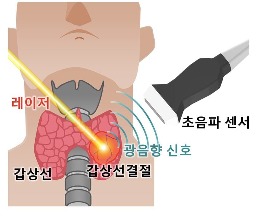
Usually, the process of diagnosing thyroid cancer starts with an ultrasound scan. If a questionable lump is found, a biopsy is carried out using a needle to obtain tissue samples. Nevertheless, ultrasound by itself is not very effective in differentiating between non-cancerous and cancerous lumps, resulting in unnecessary biopsies for benign nodules. This creates both physical and emotional stress for patients and raises issues regarding the precision of diagnosis among healthcare professionals.
A research group, backed by the POSTECH-Catholic University of Korea Institute for Biomedical Science and Technology (POGA Research Institute), created a “photoacoustic imaging (PAI)” technique. Cancerous nodules are metabolically active, leading to reduced oxygen levels. The system identifies if a nodule is benign or malignant by assessing blood oxygen saturation using faint ultrasound signals generated by red blood cells when exposed to a laser. Nonetheless, this approach had challenges in differentiating between different forms of thyroid cancer.
The research group collected information from 106 patients: 45 individuals diagnosed with papillary thyroid cancer, 32 with follicular tumors, and 29 with non-cancerous nodules. They obtained features like oxygen saturation levels, distribution asymmetry, and spectral slope from photoacoustic images and applied machine learning (AI) to create a novel diagnostic tool known as the “ATA-Photoacoustic (ATAP) score.”
The findings indicated a 97% sensitivity in identifying cancerous nodules, whereas specificity—the capacity to rule out non-cancerous nodules from further testing—rose to 38%, more than doubling the 17% seen with traditional ultrasound. This decrease in unnecessary examinations may reduce patient stress and result in lower healthcare costs.
Professor Kim Chulhong remarked, “This study is important because it integrates photoacoustic and ultrasound imaging to identify malignant conditions, such as follicular tumors, which were challenging to detect before.” Professor Park Byul-ri noted, “With further research, our goal is to improve the technology’s reliability and perform extensive clinical testing to turn it into a medical device suitable for real-world healthcare applications.”
Reference
Science Advances (2025), DOI: https://doi.org/10.1126/sciadv.ady6173


Leave a comment