
Researchers have found that changes in the shape of the brain as a person ages may signal early symptoms of dementia.
Experts are discovering that the most effective method to comprehend how the brain ages is not throughexamining individual partsbut by examining its general framework and how its various areas communicate with each other.
In a major study, scientists from Irvine,California and Tenerife, Spain, utilized brain imaging to track these structural changes. They found that as individuals grow older, the brain does not decrease in size uniformly. Rather, it undergoes distinct transformations in shape.
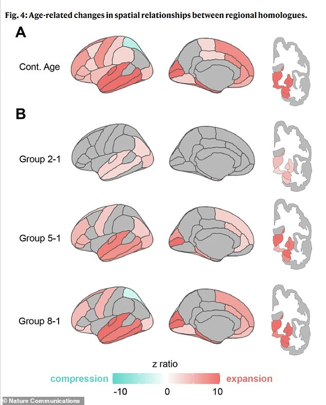
The lower sections of the brain, which manage vital processes like respiration and heart rate, along with the frontal regions, important for specific mental activities, generally grow outward.
The upper regions, essential for language abilities, and the posterior areas, responsible for visual processing and movement control, tend to fold inward.
The space between corresponding regions on the left and right sides of the brain, particularly in the frontal area, also expands.
The separation of the brain’s hemispheres physically can be a strong sign of diminished interaction and cooperation between the left and right sides of the brain. As the connection between these halves becomes weaker, the brain’s overall network operates less effectively.
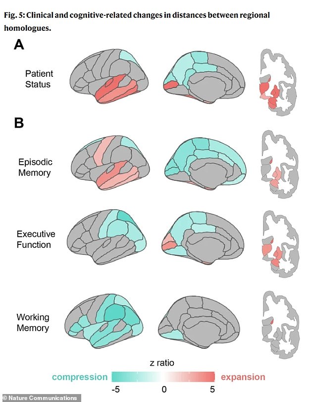
These particular structural changes, verified in several groups, were closely associated with lower cognitive abilities, including problem-solving, indicating them as a distinct physiological marker.sign of cognitive impairment.

With the US population becoming increasingly aged, the prevalence of dementia in the country is expected to increase significantly in the near future, according to current projections that indicate a rise from the present seven million cases to almost 13 million by 2060.
A typical part of aging is the slow reduction in brain size. On average, the brain decreases by approximately 0.2 percent each year after the age of 60. By the time someone turns 80, their brain could have shrunk by roughly 10 to 15 percent compared to its size in their 30s.
Dr. Niels Janssen, the lead author of the research and a professor at Universidad de La Laguna in Tenerife, Spain, stated: ‘Most investigations on brain aging concentrate on the amount of tissue lost in various areas.’
What we discovered is that the general structure of the brain changes in consistent patterns, and these changes are strongly associated with whether an individual exhibits cognitive decline.
To discover how the brain’s structure evolves over time, scientists carried out an extensive brain mapping initiative.
They began with an extensive dataset containing more than 2,600 brain scans from adults aged 30 to 97, including some individuals with dementia. They selected one large group of scans as their primary test set and a second, entirely separate group to verify their findings.
They examined the brain’s structure, including its shape and configuration, in two different methods. Initially, they studied the brain’s overall form by digitally marking 400 points on its exterior.
They subsequently calculated the distances between corresponding points on the left and right sides, generating a comprehensive map indicating areas where the brain was expanding or contracting.
Second, they calculated the distance between particular partner regions in the left and right hemispheres. Lastly, they linked these physical measurements to actual real-world functions.
By employing sophisticated metrics, they examined if these trends of growth and shrinkage were associated with the participants’ age, their results on memory and logical thinking assessments, and if they had ever received a diagnosis related to cognitive difficulties.
This enabled them to observe not only how the brain alters its form, but also how these particular changes could influence an individual’s mental capabilities.
The brain experiences a distinct and significant transformation as it ages, rather than simply decreasing in size. Certain patterns of growth and contraction were closely associated with individuals’ neurological well-being.
Memory issues have been associated with growth in areas of the temporal lobe, often referred to as the memory centers. One of the most remarkable discoveries from the study concerns the entorhinal cortex, a key center for memory located within the medial temporal lobe.
The study indicates that changes in the brain associated with aging could physically compress this delicate area against the skull’s base.
As this region is also where harmful tau protein initially accumulates in Alzheimer’s, the researchers suggested that these mechanical and gravitational forces might be an unexplored factor contributing to its high susceptibility.
Dr. Michael Yassa, one of the study’s co-authors and a neurobiologist at the University of California, Irvine, stated, “This may help clarify why the entorhinal cortex is the primary site where Alzheimer’s disease begins.”
If the aging brain is slowly changing in a manner that presses this delicate area against a hard surface, it could lead to the ideal conditions for damage to occur. Grasping this process offers us a completely new perspective on the causes of Alzheimer’s disease and the potential for early identification.
Impaired executive function, involving tasks such as planning and logical thinking, was associated with reduced volume in the parietal lobes, brain regions responsible for processing information and visual perception.
The patterns observed in individuals with clinical impairments, like a dementia diagnosis, were even more evident compared to those in healthy aging, indicating that this transformation progresses more rapidly when a disease is present.
Janssen stated, “This goes beyond simply assessing brain shrinkage. It involves observing how the brain’s structure reacts to aging and how this structure can indicate who is more prone to experience difficulties with memory and cognition.”
The finding that the brain’s structure may act as an indicator for dementia represents a major change in the way Alzheimer’s disease and other types of dementia, which lead to reduction in the hippocampus and frontal cortex, are diagnosed.
Their results were featured in the publicationNature Communications.
The weakening of specific areas of the brain might be identifiable well before significant neuronal loss takes place.
A standard MRI scan might be examined to identify this condition. A particular pattern of swelling and contraction could indicate a patient as being at high risk several years prior to memory tests revealing an obvious issue.
A neurologist can review a brain scan of a patient and detect a distinctive pattern associated with Alzheimer’s, like significant expansion in the temporal lobe combined with compression in the parietal region, as opposed to a pattern linked to a different condition, resulting in more precise diagnoses and tailored treatment strategies.
- Is your brain’s aging process in danger? Learn about the innovative assessment that could ‘effectively rule out’ dementia through a single straightforward exercise.
- Is your brain’s activity suggesting a potential future with Alzheimer’s, and might it hold the secret to early detection?
- Are indicators of decreasing cognitive function pointing towards the solution for early Alzheimer’s treatment?
- Could your stomach be signaling early signs of Alzheimer’s? A pioneering study reveals the connection between the gut and brain, offering a potential way to foresee memory loss years ahead!
- Have researchers recently revealed that your biological age could be the hidden factor behind your risk of dementia?


Leave a comment